Get Mount Fuji Sign In Tension Pneumocephalus Images. The presence of the mount fiji sign helps distinguish between pathologic subdural air causing mass effect (i.e. Tension pneumocephalus) from subdural air normally observed after sdh evacuation (i.e.
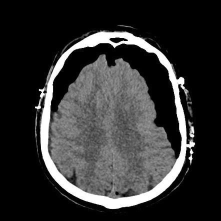
The mount fuji sign in tension pneumocephalus is described as bilateral subdural hypoattenuating air collections which cause compression and separation of the frontal lobes.1 the compressed frontal lobes and the widening of the interhemispheric space between the tips of the frontal lobes have the appearance of the silhouette of mount fuji.
2 correlation of the imaging features of tension pneumocephalus with signs of increased. Tension pneumocephalus is a surgical emergency, which needs immediate intervention in the form of decompression of the cranial cavity by a burr hole or needle aspiration. Unenhanced axial ct brain slice on brain windows demonstrating the mount fuji sign. Computed tomography imaging reveals mass effect on the ventricular system and the classic mount fuji sign, with subdural free air compressing the frontal lobes and widening the interhemispheric fissure, simulating the silhouette of mount fuji.